 |
 |
 |

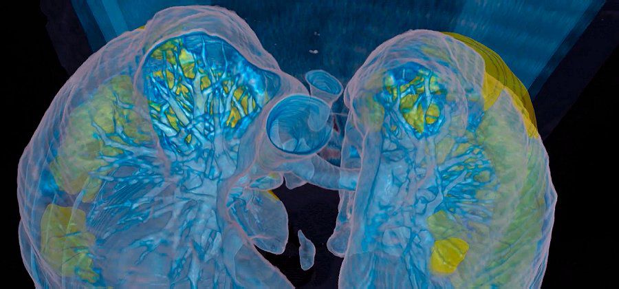
VIRTUAL REALITY IMAGE
LUNGS REACHED BY THE CORONAVRUS | |
"There is such a contrast between the abnormal infected lung and the adjacent healthy lung tissue." You don't have to be a doctor to understand these images, ”says Dr. Keith Mortman, chief of thoracic surgery at GW University Hospital. |
|
The severity of the infection highlighted The patient, close to sixty, was already in intensive care and required the help of a respirator. To better understand the degree of progress of the disease. In the video healthy tissues are represented in blue, the infected parts appear in green and yellow. The disease is diffuse and causes serious damage to both lungs. For the doctor, it is likely that long-term lung damage will remain in very seriously ill patients who may have survived the virus. About 20% of Covid-19 patients develop severe symptoms and some of them require mechanical assistance to breathe. No age class is spared. |
|
| Andrew Preston for DayNewsWorld | |
 |
|